
Why tachyarrhythmias matter for the FRCEM Final
The adult tachyarrhythmia algorithm changed a lot in the 2025 Resuscitation Council UK (RCUK) guidelines, and it was revised again in January and March 2026. Several answers that were right under the 2021 algorithm are now wrong, so it’s a good topic for the FRCEM Final.
Most people can follow an algorithm. The examiners love the small print: the decisions where things don’t go to plan, the patient is complicated, or the ECG isn’t as straightforward as you think. That’s where a consultant comes into their own. You need to:
- know the fine detail of each step of the algorithm;
- be able to make decisions from ECGs that don’t completely follow the typical pattern;
- know what to do if the initial steps don’t work;
- be aware of the complications of each treatment, and where things can go wrong at each step.
What changed in the 2025 tachyarrhythmia algorithm?
Here are the main changes from the 2021 algorithm. A previously correct answer may now appear as a distractor, and in the stress of the exam your brain may default to the answer you learnt years ago. Make sure you know the changes.
- Stable VT: give a synchronised shock under sedation or anaesthesia first. Previously, the first step was amiodarone.
- Procainamide 10–15 mg/kg is an alternative to amiodarone. It’s available in the UK but unlicensed, so amiodarone remains an option where procainamide isn’t available.
- Life-threatening features: “immediately after ROSC” is new, “syncope” now reads “syncope with severe or ongoing hypotension”, and “severe heart failure” now reads “severe heart failure with pulmonary oedema”.
- SVT with aberrancy: the algorithm separates suspected (give adenosine, then treat as VT if it fails) from known (treat as a regular narrow-complex tachycardia).
- AF with bundle branch block is no longer listed in the broad-complex part of the algorithm.
- AF: anticoagulate if it has lasted more than 24 hours (previously 48), and control the rate according to the ejection fraction.
- Torsades de pointes: magnesium 8 mmol (2 g magnesium sulfate) as before; consider isoprenaline or temporary pacing; avoid amiodarone.
- Sinus tachycardia: the algorithm now includes a reminder that it isn’t for sinus tachycardia, where you treat the cause.
The 2025 adult tachyarrhythmia algorithm
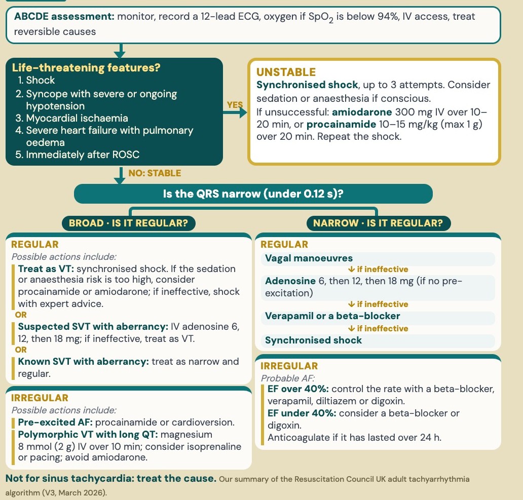
Our summary of the Resuscitation Council UK adult tachyarrhythmia algorithm (V3, March 2026). Print the original from the link at the end.
How do you assess a patient with a tachyarrhythmia?
- Airway: check it’s patent.
- Breathing: monitor the SpO2 and give oxygen if it’s below 94%. Look for pulmonary oedema.
- Circulation: monitor the ECG and blood pressure, record a 12-lead ECG and obtain IV access. Look for shock and myocardial ischaemia, and check the electrolytes on a blood gas.
- Disability: check the conscious level, and ask about syncope.
- Exposure: look for reversible causes, such as sepsis or bleeding, and check the drug history and drug chart.
The algorithm is for peri-arrest tachyarrhythmias of abnormal origin. In sinus tachycardia, treat the underlying cause: don’t try to slow the heart rate with antiarrhythmics or synchronised cardioversion. This isn’t new practice, but the 2025 algorithm now states it in a box of its own, probably as a reminder. If a patient in sepsis or bleeding has a sinus tachycardia of 150, treat the sepsis or the bleeding, not the rate: don’t give adenosine or a beta-blocker (see our sepsis guide).
What are the key features of an unstable tachycardic patient?
Look for the five life-threatening features. If you learnt the previous four years ago, they’re probably etched in your memory, so make a point of committing the changes:
- Shock: hypotension (for example, a systolic below 90 mmHg) with signs of compensation and inadequate organ perfusion.
- Syncope with severe or ongoing hypotension (previously, just “syncope”).
- Myocardial ischaemia: chest pain, or ischaemia on the 12-lead ECG, which can be silent.
- Severe heart failure with pulmonary oedema (previously, “severe heart failure”).
- Immediately after ROSC (new in 2025).
How do you treat an unstable tachycardic patient?
If any of the five features is present, give a synchronised shock, up to 3 attempts. If the patient is conscious, give careful sedation or anaesthesia, and be aware that it can cause haemodynamic deterioration (see cardioversion: the small print).
If the patient remains unstable after 3 shocks, give amiodarone 300 mg IV over 10–20 minutes or procainamide 10–15 mg/kg (maximum 1 g) over 20 minutes (new in 2025), then repeat the shock. The loading dose of amiodarone can be followed by 900 mg over 24 hours.
For the energies, synchronising and sedation, see cardioversion: the small print.
Unstable AF
Give a synchronised shock at maximum output. With a severely reduced ejection fraction, consider digoxin or amiodarone for acute rate control. With acute decompensated heart failure, avoid calcium-channel blockers, and involve cardiology before giving a beta-blocker. Here “calcium-channel blockers” means verapamil and diltiazem: amiodarone is a class III antiarrhythmic, although it has some calcium-channel-blocking effect.
In a stable patient, is the QRS broad or narrow?
If none of the five life-threatening features is present, the patient is stable. Then ask two questions:
- Is the QRS narrow (under 0.12 seconds)?
- Is it regular?
That gives four groups, each covered below: broad and regular (VT, or SVT with aberrancy), broad and irregular (pre-excited AF, torsades, or AF with bundle branch block), narrow and regular (SVT) and narrow and irregular (probable AF).
The stable part of the algorithm “does not address every possible scenario” (RCUK). The causes of a tachyarrhythmia can be many and varied, and the algorithm is a guide that’s safe, quick and easy to follow. The more expertise someone has in ECGs and cardiology, the more nuanced their management of a stable patient can be. If the patient becomes unstable at any point, treat them as unstable.
Broad and regular: VT
How to recognise VT
About 80% of broad-complex tachycardias are VT, and drugs used for SVT can harm a patient in VT (ESC). So if in doubt, treat it as VT. Look for the features that confirm it:
- AV dissociation (more QRS complexes than P waves) and capture or fusion beats: the key diagnostic features.
- A QRS over 140 ms with a right bundle branch block pattern, or over 160 ms with a left bundle branch block pattern.
- An extreme axis (−90 to ±180°).
- Negative concordance across the chest leads (all QRS complexes pointing down).
Use the history and any old ECG too: treat an older patient with a previous MI or a cardiomyopathy as being in VT until proven otherwise. A bundle branch block on the opposite side on an old ECG in sinus rhythm also points towards VT (ESC).
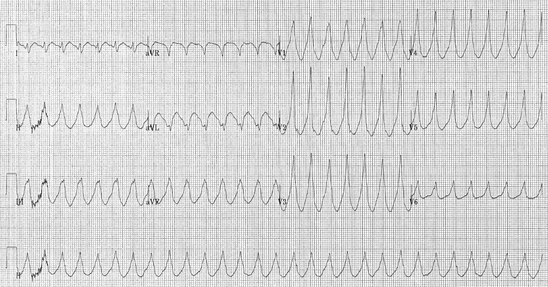
Monomorphic VT: a regular broad-complex tachycardia in every lead. Karthik Sheka, MD, via Wikimedia Commons, CC BY-SA 2.5 (shared unmodified).
How to treat stable VT: shock first
Give a synchronised shock under sedation or anaesthesia (previously, the first step was amiodarone), starting at 120–150 J.
Why shock a patient who is stable? If the patient has structural heart disease, or you don’t know whether there’s underlying heart muscle damage, untreated monomorphic VT can cause haemodynamic deterioration, so cardioversion is recommended even though they’re stable (RCUK, ERC).
If the sedation or anaesthesia risk is too high, give procainamide 10–15 mg/kg over 20 minutes (new in 2025). If it isn’t available or is contraindicated, give amiodarone 300 mg IV over 10–60 minutes, then 900 mg over 24 hours.
If the drugs don’t work, RCUK recommends synchronised shocks with expert advice. Shocking a conscious patient without sedation or anaesthesia isn’t recommended, so you need advice from anaesthetics and cardiology at this point. Know who the experts are, because the exam could test it: anaesthetics for safe sedation or anaesthesia, and cardiology for the arrhythmia itself.
Broad and regular: SVT with aberrancy
SVT with aberrancy is an SVT conducted with a bundle branch block. The causes are:
- a pre-existing bundle branch block;
- a bundle branch block that develops at the fast rate, more often right-sided;
- QRS widening from drugs, such as flecainide, or electrolyte disturbance.
How to recognise it
- On the ECG: a typical bundle branch block pattern, with none of the features of VT (no AV dissociation, capture or fusion beats, extreme axis or negative concordance). In a typical right bundle branch block, V1 shows rSR′ or rR′ and V6 has an R wave taller than the S wave. In a typical left bundle branch block, there’s no Q wave in V6 (ESC).
- The patient: typically younger, with no ischaemic or structural heart disease.
- An old ECG: the same bundle branch block in sinus rhythm makes SVT with aberrancy more likely, but see the caution below.
Don’t rely on the bundle branch block shape alone. V1 and V6 often disagree: in about a third of cases, one looks like SVT with aberrancy and the other looks like VT (ESC). If there’s any doubt, treat as VT.
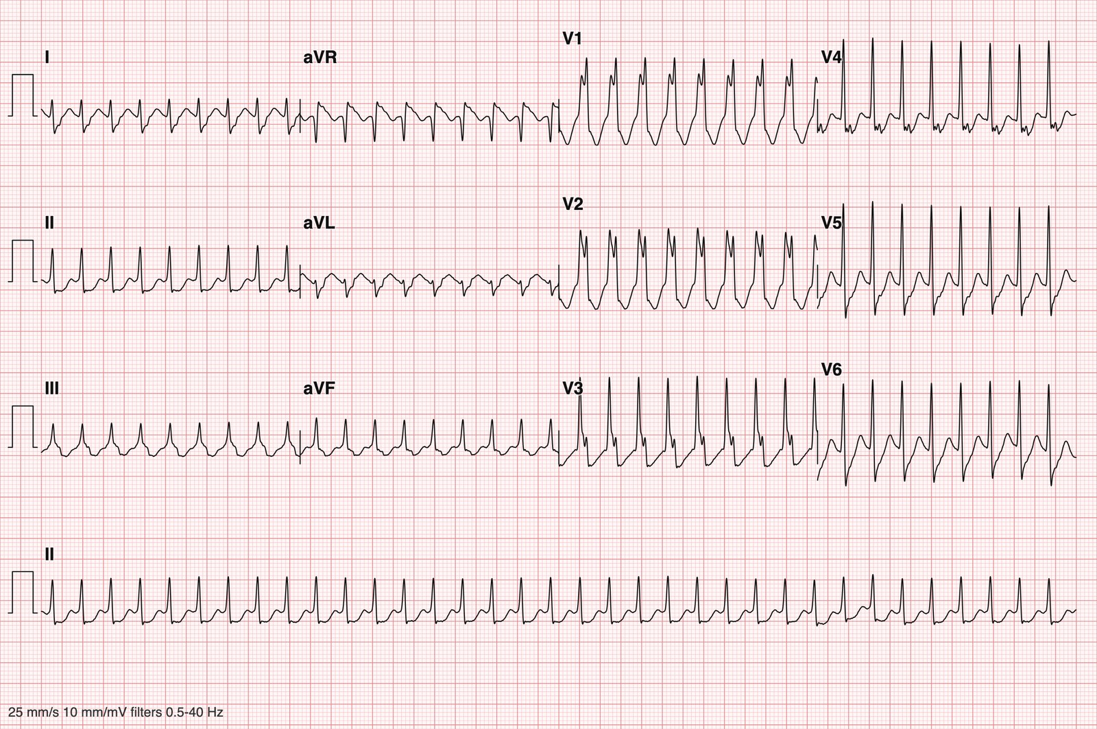
Labelled SVT with right bundle branch block: regular at about 210/min, rSR′ in V1 with the right “ear” taller, a broad S wave in lead I, a normal axis, and no AV dissociation or capture beats. Chapman–Shaoxing 12-lead ECG database (Zheng J et al., Sci Data 2020), CC0, re-plotted from the raw data.
Suspected SVT with aberrancy
Suspect it in a younger patient with no ischaemic or structural heart disease. Give IV adenosine 6 mg, then 12 mg, then consider 18 mg, as a rapid bolus into a large, proximal vein with a 12-lead ECG running. If it’s ineffective, treat as VT (new in 2025).
Known SVT with aberrancy
“Known” means a previous specific diagnosis, for example an old ECG showing the same bundle branch block during a documented SVT. It’s the combination that needs to be known: a history of SVT on its own, or a bundle branch block on an old ECG in sinus rhythm, makes it suspected, not known. Treat known SVT with aberrancy as a regular narrow-complex tachycardia, starting with vagal manoeuvres. The third step there includes verapamil, so be sure of the diagnosis first: in VT, verapamil can cause severe hypotension and collapse, so avoid it in any broad-complex tachycardia that could be VT (ESC).
Don’t assume an old ECG is the patient’s normal. It may have been recorded when they presented with a previous SVT, VT or another illness. Rely on it only if a confirmed diagnosis goes with it.
There are three broad, irregular rhythms to tell apart: pre-excited AF, torsades de pointes and AF with bundle branch block. Look at the ECG and the history together.
Broad and irregular: pre-excited AF
AF in a patient with an accessory pathway (Wolff–Parkinson–White). It’s now listed in the algorithm (new in 2025).
How to recognise it
- During the tachycardia: it’s fast, broad and irregular, with a very rapid rate, often over 200 a minute (faster than the AV node usually allows in AF), and a QRS shape and width that vary from beat to beat (ESC).
- On an old ECG in sinus rhythm, or after cardioversion: the WPW pattern of a short PR interval (120 ms or less), a delta wave (a slurred upstroke of the QRS) and a broad QRS (ESC).
- The patient: often young, and may know about their WPW or have had palpitations before.
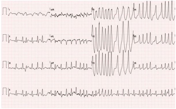
Pre-excited AF: fast, broad and irregular, with the QRS shape varying from beat to beat. Kesler K, Lahham S. West J Emerg Med 2016, CC BY 4.0 (shared unmodified).
How to treat it
- Treat with procainamide 10–15 mg/kg (maximum 1 g) IV over 20 minutes, or a synchronised shock at maximum output.
- Avoid AV nodal blockers: adenosine, verapamil, diltiazem, beta-blockers and digoxin. With the AV node blocked, the AF conducts down the accessory pathway, which has a shorter refractory period, so the ventricular rate rises, with a risk of VF.
- Avoid amiodarone too: it can enhance conduction down the pathway, and VF has been reported (ESC).
Broad and irregular: torsades de pointes
How to recognise it
- On the ECG: a polymorphic VT in which the QRS axis twists around the baseline. It often comes in runs.
- Before it starts, or between runs: look for a long QT interval.
- The history: the drug history or a condition in the stem may give you the clue.
- QT-prolonging drugs, for example sotalol and amiodarone, macrolide antibiotics (clarithromycin, erythromycin), antipsychotics (haloperidol), citalopram, methadone and ondansetron.
- Conditions: a low potassium, magnesium or calcium, a slow heart rate, and congenital long QT syndrome.
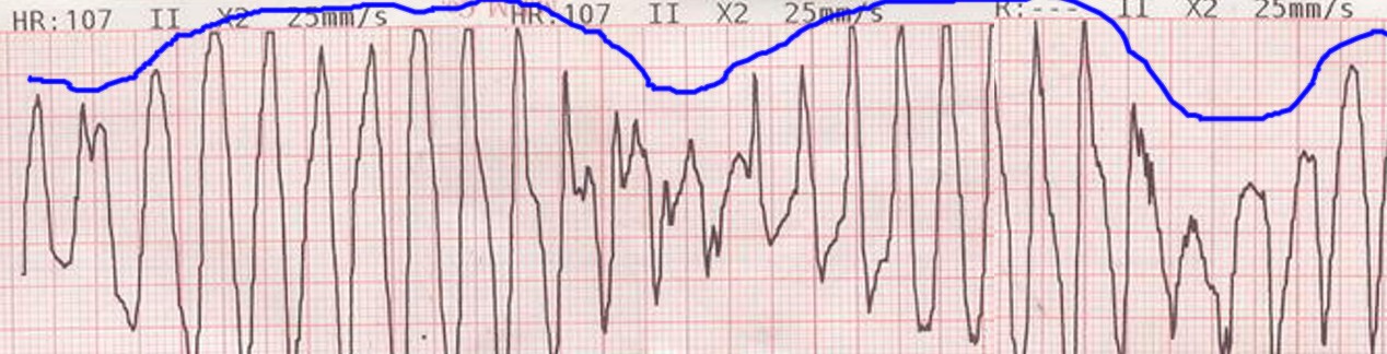
Torsades de pointes. Panthro, via Wikimedia Commons, public domain.
How to treat it
- Give magnesium 8 mmol (2 g magnesium sulfate) IV over 10 minutes, repeated once if needed. It can suppress torsades even when the serum magnesium is normal.
- Look for and remove the precipitating factors, such as QT-prolonging drugs, and correct the potassium.
- Consider isoprenaline or temporary pacing (new in 2025): increasing the heart rate can suppress it.
- Avoid amiodarone (new in 2025), which prolongs the QT.
What about AF with bundle branch block?
How to recognise it
- On the ECG: broad and irregularly irregular, with no P waves, but the QRS shape stays the same every beat, with a typical bundle branch block pattern. The rate is usually slower than in pre-excited AF.
- On an old ECG: the same bundle branch block may be there in sinus rhythm.
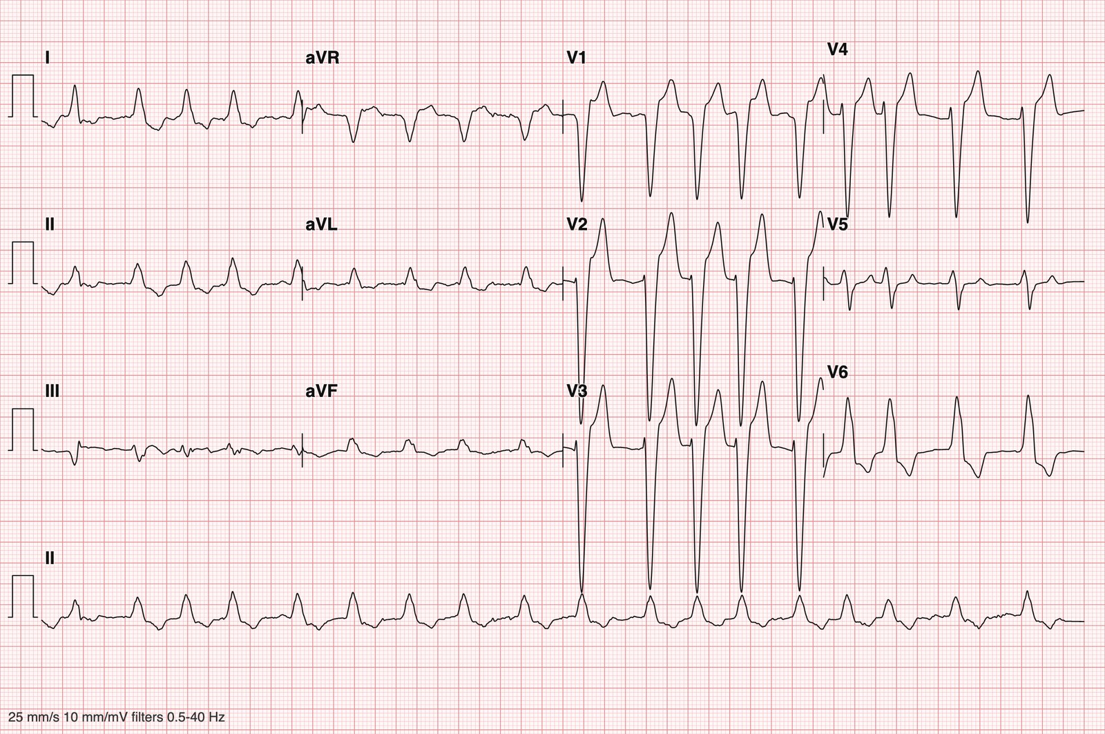
Labelled AF with left bundle branch block: irregular, with the same broad left bundle branch block shape on every beat. PTB-XL, Wagner P et al., PhysioNet 2022, doi:10.13026/kfzx-aw45, CC BY 4.0. Re-plotted from the raw data, filtered 0.5–40 Hz.
How to treat it
It’s no longer listed in the broad-complex part of the algorithm (previously, “treat as for irregular narrow complex”). Treat it the same as narrow-complex AF: control the rate by ejection fraction and anticoagulate (see narrow and irregular: probable AF).
Narrow and regular: SVT
How to recognise it
- On the ECG: a regular narrow-complex tachycardia. In AVNRT, the commonest type, the P waves are usually hidden in the QRS or seen just after it (ESC).
- A rate of about 150: look for atrial flutter with 2:1 block, with sawtooth flutter waves in the inferior leads (ESC).
- Sinus tachycardia has a normal P wave before every QRS: look for the cause.
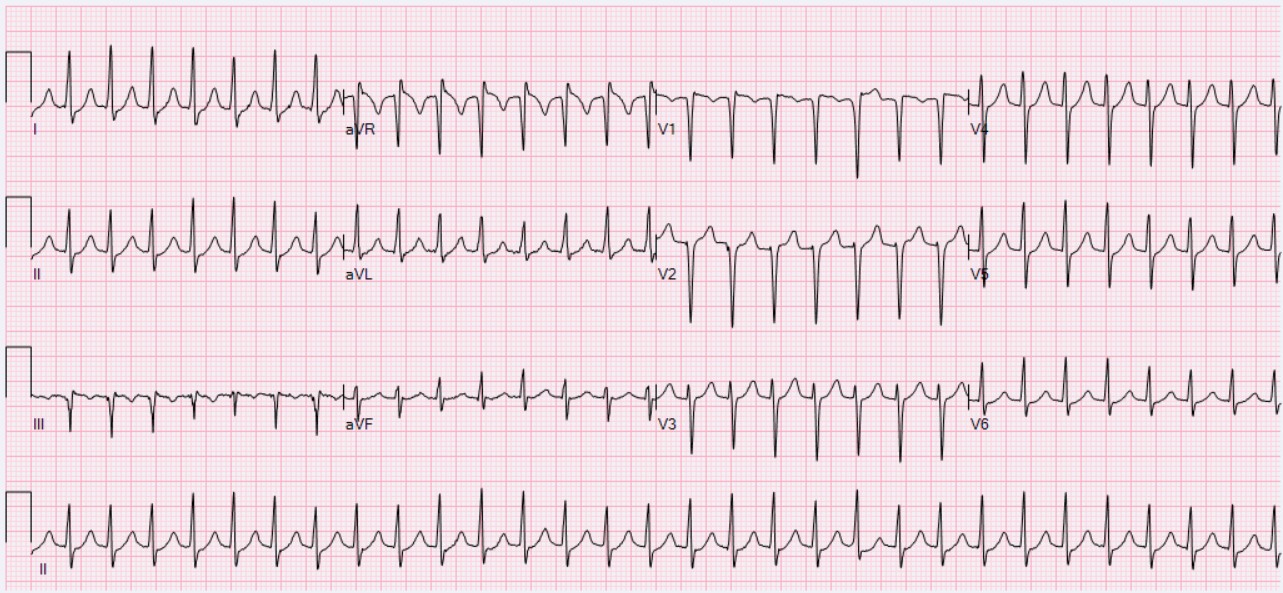
AVNRT: a regular narrow-complex tachycardia at 181/min. Ewingdo, via Wikimedia Commons, CC BY-SA 4.0 (shared unmodified).
How to treat it, step 1: vagal manoeuvres
Vagal manoeuvres stimulate the vagus nerve, which slows conduction through the AV node. The modified Valsalva manoeuvre converted 43% of patients, compared with 17% for the standard Valsalva, in the REVERT trial.
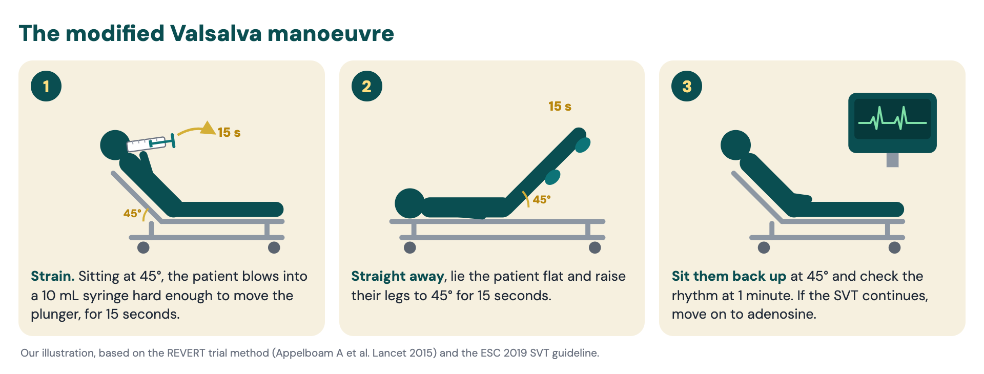
Our illustration, based on the REVERT trial method (Appelboam A et al. Lancet 2015) and the ESC 2019 SVT guideline.
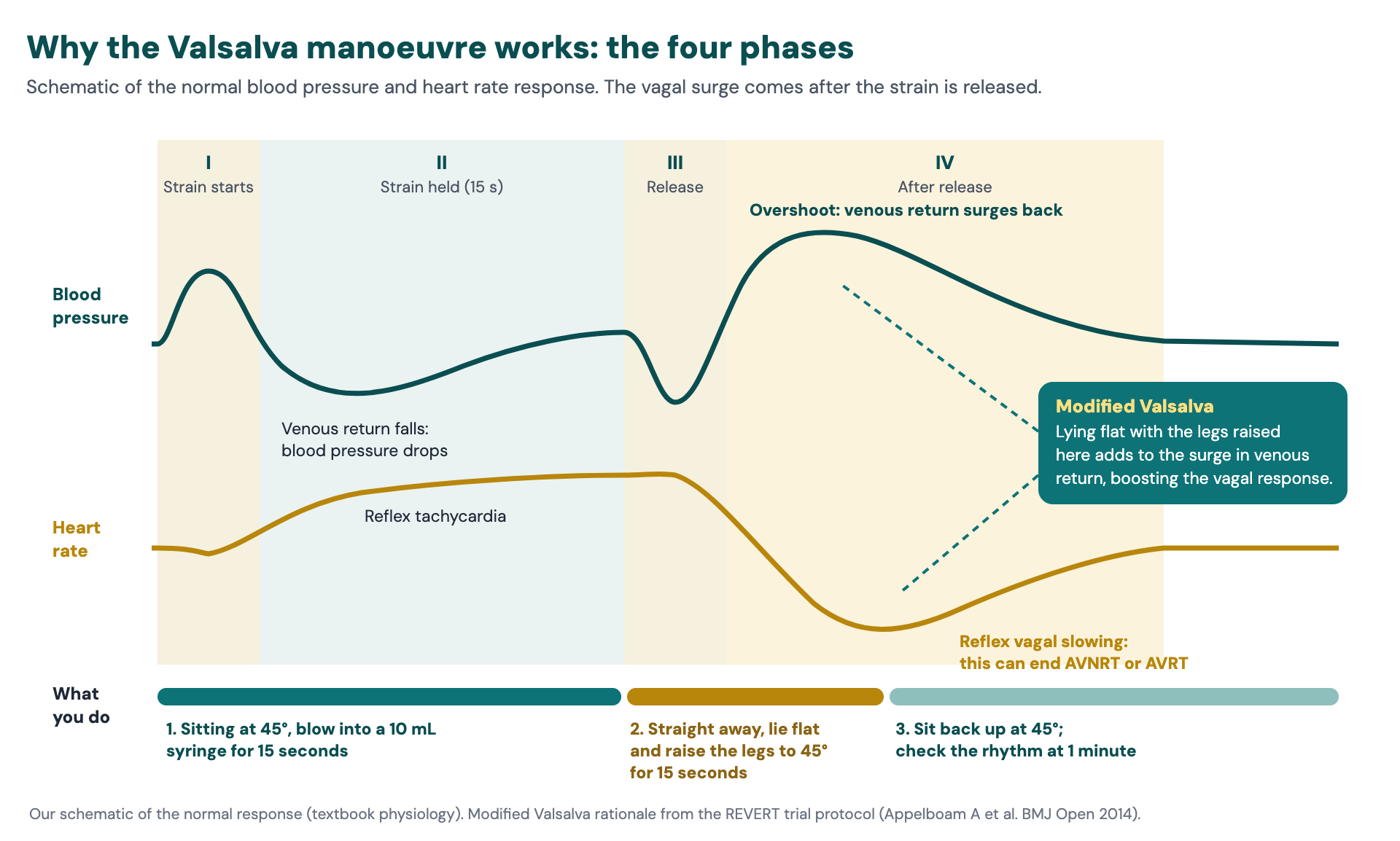
Our schematic of the normal response (textbook physiology). The vagal slowing comes after the strain is released; lying flat with the legs raised adds to the surge in venous return at that point (REVERT trial protocol, Appelboam A et al. BMJ Open 2014).
In late pregnancy: when the woman lies flat, the uterus compresses the inferior vena cava and reduces venous return, which is the opposite of what the leg raise is for. Use manual uterine displacement during the leg-raise phase.
Other vagal manoeuvres aren’t ruled out, but the modified Valsalva is the most effective (ESC):
- Carotid sinus massage: on one side only, for no more than 5 seconds, with the neck extended, the head turned away and the patient monitored. Avoid it after a previous TIA or stroke, or if there’s a carotid bruit (ESC).
- Facial immersion in cold water, or forceful coughing: rarely used now (ESC).
Step 2: adenosine
If vagal manoeuvres fail, and there’s no pre-excitation, give adenosine 6 mg as a rapid IV bolus, then 12 mg, then 18 mg. Give it into a large, proximal vein with a flush, with a continuous 12-lead ECG running.
- If adenosine terminates the rhythm, it was AVRT or AVNRT.
- If it doesn’t, look at the 12-lead ECG recorded during the dose. Adenosine blocks the AV node for a few seconds, and you may see the underlying atrial rhythm, such as flutter. When adenosine “doesn’t work”, you may still have the diagnosis on the ECG.
- Use clinical judgement with a history of asthma: adenosine can rarely provoke severe bronchospasm.
Step 3: verapamil or a beta-blocker, then a shock
If adenosine fails, give verapamil or a beta-blocker. If that fails, give a synchronised shock, starting at 70–120 J.
Narrow and irregular: probable AF
How to recognise it
An irregularly irregular narrow-complex tachycardia, with no P waves. The algorithm calls it “probable AF” because it’s usually AF, but check the 12-lead ECG for atrial flutter with variable block, which has flutter waves.
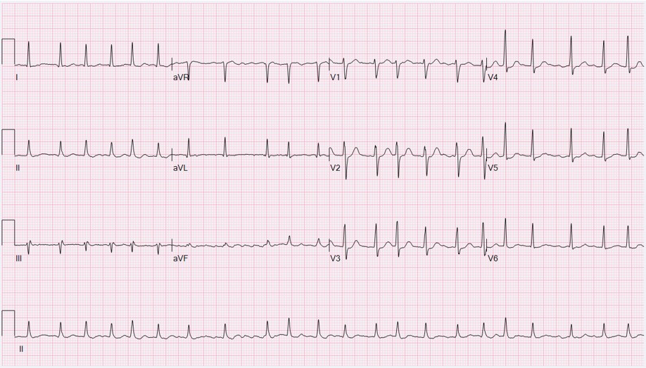
AF with a fast ventricular response: irregularly irregular, with no P waves. Ewingdo, via Wikimedia Commons, CC BY-SA 4.0 (shared unmodified).
How to treat it
What the RCUK algorithm recommends
- Rate control, chosen by the ejection fraction. With an EF over 40%, a beta-blocker, verapamil, diltiazem or digoxin (previously, a beta-blocker). With an EF under 40%, consider a beta-blocker or digoxin, aiming for a heart rate under 110/min (previously, digoxin or amiodarone if there was heart failure).
- Anticoagulation if the AF has lasted more than 24 hours (previously, 48 hours).
Rate or rhythm control?
Within 24 hours of onset, you can control either the rate or the rhythm. The 24-hour cut-off is new: RCUK (2025) and the European Society of Cardiology (ESC, 2024) have moved it from 48 hours. NICE (NG196) still uses 48, but its acute AF recommendations date from 2014, so go with the more recent guidance.
The RCUK algorithm is a quick, easy-to-use reference for anyone, so its treatment is the failsafe one: rate control. That’s also a sound choice in a stable patient (ESC). About two-thirds of patients revert on their own with rate control alone, and electrical cardioversion means sedation and its risks.
Rhythm control is more nuanced, which is why it’s hard to fit into a simple algorithm. If the onset is definitely within 24 hours, the patient would benefit from a quick solution and you have the resources, you could choose electrical cardioversion, which is quick, or drug cardioversion, which is slower. If a question is looking for one answer over the other, the stem will give you the clues.
Over 24 hours, or if the onset is uncertain, use rate control.
Rhythm control
- When: within 24 hours of onset, without anticoagulating first. After 24 hours, or if the onset is uncertain, only after at least 3 weeks of therapeutic anticoagulation, or once a transoesophageal echo (TOE) has excluded a thrombus.
- How: electrical cardioversion under sedation (see cardioversion: the small print), or drugs. With no structural or ischaemic heart disease, give flecainide or amiodarone; with structural heart disease, amiodarone. Don’t use magnesium or a calcium-channel blocker to cardiovert (NICE).
- Afterwards: continue anticoagulation for at least 4 weeks, then long term according to the stroke risk (ESC).
Anticoagulation
If the patient isn’t anticoagulated, start heparin at presentation unless it’s contraindicated (NICE), then continue according to the stroke risk.
Cardioversion: the small print
Synchronising
- Time the shock to the R wave: a shock on the T wave can cause VF.
- Check the sync marker is on every R wave. If it isn’t, change the lead or the amplitude. This can be difficult in VT, with its broad, varied complexes.
- Check sync is still on after every shock, and keep the shock button pressed until the shock is delivered.
- In unstable VT, if synchronising fails, give unsynchronised shocks rather than delay. VF and pulseless VT need unsynchronised shocks.
- For atrial rhythms, use antero-posterior pads if possible.
Energies (unchanged)
- AF: maximum output from the first shock, rather than escalating.
- Flutter and SVT: 70–120 J, then increase with each shock.
- VT with a pulse: 120–150 J, then consider increasing.
Sedation
With a poor cardiac output, sedatives take longer to work and smaller doses have a bigger effect, including hypotension. Titrate slowly, involve anaesthetics or ICU, and consider when the patient last ate (see our procedural sedation guide).
If a junior colleague is cardioverting, check what they’re doing and teach as you go: that sync is on, the energy is right and the sedation plan is safe.
What if the arrhythmia persists or treatment fails?
- Recurrent or persistent arrhythmia: persistent arrhythmias need careful evaluation. They’re often linked to structural heart disease, and may point to unresolved myocardial ischaemia. Look for the cause: the patient may need PCI rather than another antiarrhythmic.
- Shocks not working: if the patient remains unstable after 3 shocks, give procainamide or amiodarone, then shock again.
- Read the history for the cause: a previous MI or cardiomyopathy (think VT); known WPW (pre-excited AF); QT-prolonging drugs or a low potassium (torsades).
- Specialty input: you decide when to involve cardiology, anaesthetics or ICU, depending on how complicated the patient is and how they’re responding to your treatment. For example, cardiology for VT that keeps recurring with ischaemia, or anaesthetics when sedation for cardioversion is high risk.
What are the exam tips for tachyarrhythmias?
The examiners love the small print. Most people can follow an algorithm, so expect questions on what to do when things don’t go to plan, the patient is complicated, or the ECG isn’t as straightforward as you think.
A previously correct answer may appear as a distractor. In the stress of the exam, your brain may default to the answer you learnt years ago, so make sure you know the changes.
In the grey areas, read the stem carefully. If the exam wants you to choose one answer over another, the stem will steer you there with clues.
Further reading
For recognising these rhythms on the ECG, see our ECG guide for the FRCEM Final. If two options both look right, see when two answers look right.
Read the guidelines in full
- Print the RCUK adult tachyarrhythmia algorithm (V3, March 2026), and read the RCUK adult advanced life support guidelines (2025), the RCUK ALS FAQs and the RCUK adult quick reference handbook (card 2-2, compromised tachycardia).
- European Resuscitation Council guidelines 2025: adult advanced life support.
- ESC guidelines for the management of supraventricular tachycardia (2019).
- ESC guidelines for the management of atrial fibrillation (2024).
- NICE NG196, Atrial fibrillation: diagnosis and management (2021).
- The REVERT trial: postural modification to the standard Valsalva manoeuvre (Lancet, 2015).
Drug doses are for reference only — confirm against the British National Formulary (BNF) and the current guidelines.
Practise the next step
Every question in our FRCEM Final SBA bank comes with a detailed explanation and links to the guidelines, so you see why the answer is right as soon as you’ve chosen it. If you want help finding where your marks are going, I also offer 1:1 coaching on technique, the question stems and your specific gaps.
Try 10 free questions →Sign me up →About Dr Vickie Fletcher